
The neck carries the vital arteries transmitting blood to the brain, head and face. It also transmits the veins which drain the deoxygenated blood from the brain, head and face and return it to the right side of the heart. The neck also contains the cervical spinal cord, as well as some important cranial nerves. Some of these structures travel together in a fascial (connective tissue) sheath called the carotid sheath. The carotid sheath carries the common and internal carotid arteries, the internal jugular vein, the vagus nerve (cranial nerve X), some lymph nodes, carotid periarterial plexuses and the carotid sinus nerve.
Arterial Anatomy
The main artery in the neck is the common carotid artery, which divides at the upper border of the thyroid cartilage of the larynx (C4). The left common carotid artery branches directly off the aortic arch and extends into the neck. The right common carotid artery has a different initial course. It is a branch of the brachiocephalic trunk. The brachiocephalic trunk is the first branch of the aortic arch and it bifurcates into the right subclavian artery and the right common carotid artery. Both common carotid arteries ascend in the neck lying medially in the carotid sheath.
Near the bifurcation of the common carotid arteries, there are two important receptors, the carotid sinus and the carotid body. The carotid sinus is a baroreceptor which senses the pressure in the carotid artery system and transmits information to the brain about the blood pressure in order to maintain blood pressure homeostasis. It is located at the beginning of the internal carotid artery as a small dilatation. The carotid body is a small lump of tissue that lies on the medial side of the common carotid bifurcation. Its role is to monitor the levels of oxygen in the blood; it relays information about blood oxygen to the brain. In accordance with the changes sensed by the carotid body, the brain responds by changing the rate of breathing.
The internal carotid artery is the main supplier of blood to the brain. It arises from the common carotid artery in the neck, and courses posterior to the external carotid artery. The internal carotid artery does give off any branches in the neck. It ascends into the cranial cavity through the carotid canals in the petrous part of the temporal bone.
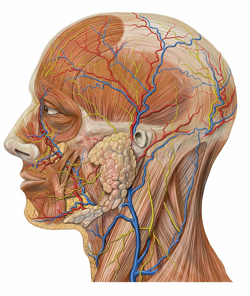
The external carotid artery is the other branch of the common carotid artery after it bifurcates at the upper border of the thyroid cartilage. It lies anterior to the internal carotid artery. It ascends in the neck and enters the parotid gland on the face, where it divides into the maxillary artery and the superficial temporal arteries. The external carotid supplies parts of the neck, face and scalp primarily. However, the middle meningeal artery is a branch which supplies the dura mater; the tough, outer meningeal covering of the brain. Unlike the internal carotid artery, the external carotid gives off a number of branches in the neck. Close to the bifurcation, the superior thyroid artery branches from the external carotid. It travels in an antero-inferior direction as it descends to supply the thyroid gland. The superior laryngeal artery branches off the superior thyroid artery and supplies blood to the larynx. The superior thyroid artery also supplies blood to a number of neck muscles.
Another branch which arises close to the bifurcation is the ascending pharyngeal artery. It arises from the posterior aspect of the external carotid artery and courses upwards between the internal and external carotid arteries on the pharynx (throat), which it supplies. It also supplies blood to the deep prevertebral muscles and parts of the middle ear.
The next branch from the external carotid artery is the lingual artery, which arises close to the middle pharyngeal constrictor muscle. It supplies blood to parts of the tongue, and gives rise to the sublingual artery and the deep lingual artery. From its origin, it passes upwards and anteriorly. It travels deep to the hypoglossal nerve (the twelfth cranial nerve) for part of its course.
The facial artery is an anterior branch of the external carotid artery. It usually arises above the lingual artery, but in some people, these arteries share a common origin. The facial artery gives off two important branches in the neck; the ascending palatine artery and the tonsillar artery, which supply the tonsils, the soft palate and nearby structures. It then travels underneath muscles in the upper neck and lower jaw, and pierces the submandibular salivary gland, which it supplies. It travels along the floor of the mouth, giving off the submental artery. It then passes over the body of the mandible and enters the face, where it supplies the muscles of facial expression.
The occipital branch arises from the external carotid artery at almost the same level as the facial artery; except it arises from the posterior aspect. It courses posteriorly, crossing the anterior aspect of the internal carotid artery and courses close to the posterior belly of the digastric muscle for part of its course. It ascends towards the head and passes in a space between the transverse process of C1 and the mastoid process of the temporal bone. It gives off branches to the sternocleidomastoid muscle, the auricle, mastoid process and the occipital bone.
The next branch is the small posterior auricular artery. It ascends in a posterior direction and travels underneath the parotid salivary gland towards the ear, where it then passes between the external acoustic meatus, auricle of the ear and the mastoid process. It supplies the parotid gland, some muscles and the auricle.
The two terminal branches of the external carotid artery are the superficial temporal artery and the maxillary artery. The external artery travels to the parotid gland, where it bifurcates into the superficial temporal and maxillary arteries. From the parotid gland, the maxillary artery runs forward over the ramus of the mandible and enters a space known as the pterygopalatine fossa. The maxillary artery has a number of branches which supply deep facial structures. The superficial temporal artery ascends through the parotid gland and travels over the zygomatic arch and anterior to the auricle of the ear. It then divides into two branches, the frontal branch and the parietal branch.
Another important artery in the neck is the vertebral artery. The vertebral artery is a branch of the subclavian artery, which is the main artery to the upper limb. After branching from the subclavian artery, the vertebral artery travels upwards in a space between the scalene muscles and the longus capitis and longus colli muscles. It then enters the transverse foramen of C6 and then ascends through the transverse foramina of C6 all the way through to C1. From here, the vertebral artery passes through a groove on C1 and enters the cranium through the foramen magnum; through which the spinal cord passes. Once inside the cranium, the vertebral artery gives branches to the spinal cord, cerebellum and the medulla oblongata. At the pons of the brainstem, the two vertebral arteries unite to form the basilar artery, which supplies blood to the brain. The basilar artery is part of the Circle of Willis, which is an anastomosing network of vessels which ensure a constant blood supply to the brain.
Venous Anatomy
The largest vein in the neck is usually the internal jugular vein, which drains blood from the brain, neck muscles, face and organs of the neck. The internal jugular vein commences at the jugular foramen, and is the direct continuation of the sigmoid sinus, which is a large vein draining blood from the vein. The internal jugular vein then descends in the neck as part of the carotid sheath, where it lies laterally. It passes under the sternocleidomastoid muscle for part of its course. It unites with the subclavian vein to form the brachiocephalic vein. At the inferior end of the internal jugular vein there is a valve which prevents the retrograde flow of blood.
The drainage of other veins into the internal jugular vein is highly variable. Roughly at the level of the hyoid bone, the facial vein empties into the internal jugular vein. Prior to draining into the internal jugular vein, the facial vein often receives the superior thyroid vein and the lingual and sublingual vein. However, in other people, the lingual vein (which drains blood from the tongue) empties into the internal jugular vein at approximately the level where the lingual artery arises from the external carotid artery. A venous plexus exists on the wall of the pharynx, from which the pharyngeal vein emerges. The pharyngeal vein tends to empty into the internal jugular vein at approximately the level of the angle of the mandible. The internal jugular vein also receives the inferior and middle thyroid veins. The internal jugular vein terminates by uniting with the subclavian vein to form the brachiocephalic vein.
The other key vein in the neck is the external jugular vein. The external jugular vein forms close to the angle of the mandible and near the lowermost point of the auricle of the ear. It is formed by the union of the posterior auricular vein and the posterior division of the retromandibular vein. It passes diagonally across the sternocleidomastoid muscle. At the base of the neck, the external jugular vein receives the transverse cervical vein and the suprascapular vein. It also receives the anterior jugular vein, which begins usually at the level of the hyoid bone by the union of the superficial submandibular veins. It then descends quite superficially in the anterior neck and then travels laterally in the root of the neck to drain into the external jugular vein. The external jugular vein terminates by draining into the subclavian vein.
The post Vascular Anatomy of the Neck appeared first on ENT Clinic Sydney.